![]()
|
|
| People | Research |
|
Aptamer-based cell
manipulation In addition to targeting biomolecules, aptamers can also selectively target cells (via membrane surface-marker binding), and as such can be of great interest for clinical diagnostic and therapeutic applications. For example, aptamers have been used to harvest stem cells for tissue engineering, and to distinguish malignant and healthy cells for detection and targeted treatment of cancer. Recognizing this potential of aptamers, we have initiated the development of microfluidic platforms that exploit aptamers for manipulation, isolation and detection of cells. We have thus far focused on developing a microfluidic system for continuous-flow, magnetically controlled incubation, capture and isolation of cells and microparticles. The system overcomes limitations of existing devices in prolonged off-chip incubation, slow batch-mode operation and complicated fabrication. It consists of an active-mixing enhanced incubator (for cell/microparticle capture) and a magnetic fractionation-based separator. The incubator features a serpentine-shaped channel design that judiciously exploits an externally applied magnetic field to accomplish effective mixing and capture of target cells or particles by surface-functionalized magnetic beads. The separator (with zero dead volume) is integrated immediately downstream of the incubator; and utilizes the same external magnetic field to isolate the captured cells or particles into a lateral buffer stream. Experimental results have demonstrated that the microfluidic system offers high separation efficiency (>90%). Ongoing work involves incorporation of aptamers within the microfluidic system for capture, enrichment and isolation of cells. 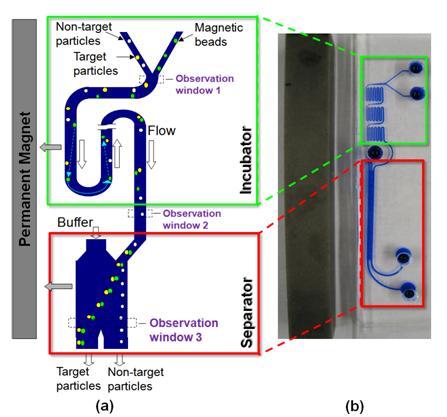 Microfluidic system for continuous-flow, magnetically controlled capture and separation of microparticles. (a) Schematic of configuration and principle of the chip, which consists of a serially connected incubator and separator placed next to a bar-shaped permanent magnet. With judiciously designed channel geometry, the chip exploits the synergy effects of laminar hydrodynamic flow and magnetic force to promote capture of target particles by surface-functionalized magnetic beads through enhanced mixing, and to selectively separate target particle-bead complexes from non-target particles. (b) A fabricated chip (with ink solution filled in the channels for enhanced clarity). 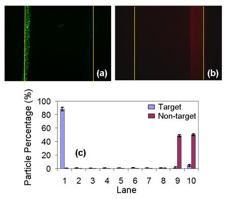 Distributions of target and non-target particles across channel width at the exit of the separator with 1:10 mixing ratio of the target vs. non-target particles. (a) Fluorescent image of the target particles. (b) Fluorescent image of the non-target particles. The yellow lines mark the channel contours. (c) Distribution of target and non-target particles in transverse lanes across the channel width, which is divided into 10 identical lanes for analysis purposes. Results show that 91.1% and 99.3% of the target and non-target particles are collected at their respective outlets, which demonstrates that the integrated device is highly efficient in capturing and isolating target particles.
|